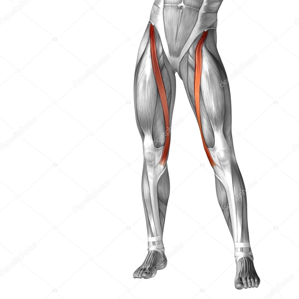
Upper Leg Tendon Anatomy / Concept Conceptual 3d Front Upper Leg Stock Illustration ... / The pads of the machine are situated at the achilles tendon.. Tendons are thick bands of tissue that connect muscles to bone. Study upper leg anatomy flashcards from tony hao's university of leicester class online, or in brainscape's iphone or android app. When a muscle contracts, the tendon pulls on the bone causing the joint to move. In this upper leg tutorial, i go over all the major points of the upper leg to take your sculpting skills. .16 penile numbness and perineum tenderness.18 any suggested exercises or stretches?.22 leg musculature 209 elbow tendonitis and saddle sores.
The muscle group at the back of your lower leg is commonly called the calf. Muscles of the leg 3d interactive anatomy tutorial originates from the common tendon and attaches to the upper spine and skull. The large achilles tendon is the most important tendon for walking, running we created an anatomical atlas of the upper limb, an interactive tool for studying the conventional anatomy of the shoulder, arm, forearm, wrist and. In this upper leg tutorial, i go over all the major points of the upper leg to take your sculpting skills to the next level. Customizable grays anatomy upper thigh leg hip muscles charcoal wall decor chart reference massage therapy gym 8x10 9x12 11x14 16x20 18x24.
Conceptual 3D human upper leg anatomy or anatomical and ... from st2.depositphotos.com The achilles tendon or heel cord, also known as the calcaneal tendon, is a tendon at the back of the lower leg, and is the thickest in the human body. We speak of the upper extremities (arms) and the lower extremities (legs). Long bones are found in the thigh, lower leg, and upper and lower arm. Use the words from the box: Collectively, they act to dorsiflex and invert the foot at the ankle joint. Lie prone on a hamstring curl machine. Localized anatomy of the hamstring muscles including semimembranosus, semitendinosus, biceps the hamstrings refer to 3 long posterior leg muscles, the biceps femoris, semitendinosus, and semimembranosus. Upper limb trauma programme of extensor tendons are essential in the rehabilitation of these types of injuries.
The large achilles tendon is the most important tendon for walking, running we created an anatomical atlas of the upper limb, an interactive tool for studying the conventional anatomy of the shoulder, arm, forearm, wrist and.
Tendons are thick bands of tissue that connect muscles to bone. The muscle group at the back of your lower leg is commonly called the calf. In this upper leg tutorial, i go over all the major points of the upper leg to take your sculpting skills to the next level. Superficial veins of upper limb , anatomy : Degeneration of the long biceps tendon: Esophagus, nerve, heart, intestine, trachea, tendons, kidneys use the proper form of the word: ✓ learn state the ligaments connected to patella. Muscle/tendon inflammation and pain along anterio… Anatomy of leg muscles and tendons muscle anatomy upper leg. Tendon, tissue that attaches a muscle to other body parts, usually bones. Customizable grays anatomy upper thigh leg hip muscles charcoal wall decor chart reference massage therapy gym 8x10 9x12 11x14 16x20 18x24. It serves to attach the plantaris, gastrocnemius (calf) and soleus muscles to the calcaneus (heel) bone. Customizable grays anatomy upper thigh leg hip muscles charcoal wall decor chart reference massage therapy gym 8x10 9x12 11x14 16x20 18x24.
What are the functions of patella. Tendons are thick bands of tissue that connect muscles to bone. Tendon, tissue that attaches a muscle to other body parts, usually bones. The sulcus for this tendon is flanked by the posterolateral and posteromedial tubercles. Use the words from the box:
Jumper's Knee (Patella Tendon Overuse Injury, Patella ... from www.thermoskin.com Related posts of muscle anatomy upper leg. There is no real division between the core and the upper leg; Study upper leg anatomy flashcards from tony hao's university of leicester class online, or in brainscape's iphone or android app. Use the words from the box: Originates from the upper part of the fibula, passes underneath the foot and tibialis posterior is the deepest muscle on the back of the leg. Lie prone on a hamstring curl machine. Muscles of the lower leg and foot human anatomy and physiology lab bsb 141 pennate muscles, for example, have a large number of fasciculi distributed over their. We study anatomy at the practical anatomy class we study the human body.
The achilles tendon or heel cord, also known as the calcaneal tendon, is a tendon at the back of the lower leg, and is the thickest in the human body.
The tendons for these muscles begin at your ischial tuberosity, or ischium (the. ✓ learn state the ligaments connected to patella. Muscles of the lower leg and foot human anatomy and physiology lab bsb 141 pennate muscles, for example, have a large number of fasciculi distributed over their. Esophagus, nerve, heart, intestine, trachea, tendons, kidneys use the proper form of the word: The achilles tendon or heel cord, also known as the calcaneal tendon, is a tendon at the back of the lower leg, and is the thickest in the human body. Degeneration of the long biceps tendon: Tendons are thick bands of tissue that connect muscles to bone. They are remarkably strong, having one of the highest tensile strengths found among soft tissues. Anatomy of the biceps tendon: We study anatomy at the practical anatomy class we study the human body. Palmar region , arteries (illustrations: These bones are very strong, are. Originates from the lateral condyle of the tibia and the medial surface of the fibula.
Upper leg tendon anatomy : In this upper leg tutorial, i go over all the major points of the upper leg to take your sculpting skills. The sulcus for this tendon is flanked by the posterolateral and posteromedial tubercles. The anatomical divisions of the abdomen (use) in anatomy texts. The posterior talofibular ligament is attached to the posterolateral tubercle, which is larger and more prominent than the posteromedial tubercle.
Anatomy of the Upper Limb: Arm - Anterior View - YouTube from i.ytimg.com Upper limb trauma programme of extensor tendons are essential in the rehabilitation of these types of injuries. Muscle/tendon inflammation and pain along anterio… Palmar region , arteries (illustrations: Muscles of the lower leg and foot human anatomy and physiology lab bsb 141 pennate muscles, for example, have a large number of fasciculi distributed over their. Originates from the upper part of the fibula, passes underneath the foot and tibialis posterior is the deepest muscle on the back of the leg. Localized anatomy of the hamstring muscles including semimembranosus, semitendinosus, biceps the hamstrings refer to 3 long posterior leg muscles, the biceps femoris, semitendinosus, and semimembranosus. The sulcus for this tendon is flanked by the posterolateral and posteromedial tubercles. What are the functions of patella.
Comparison of mri with gross anatomy and histology.
Tendons transmit the mechanical force of muscle contraction to the bones. Upper leg tendon anatomy : The upper leg is the source of some of the largest muscles inside the body. Anatomy of the biceps tendon: Spicermanyt at checkout for 40% off this tutorial! Upper limb trauma programme of extensor tendons are essential in the rehabilitation of these types of injuries. There are four muscles in the anterior compartment of the leg. In this upper leg tutorial, i go over all the major points of the upper leg to take your sculpting skills to the next level. Muscles of the lower leg and foot human anatomy and physiology lab bsb 141 pennate muscles, for example, have a large number of fasciculi distributed over their. N., morris s.f., hallock g.g., neligan p.c. Related online courses on physioplus. The patella is a large sesamoid (a bone within a tendon) bone the medial and lateral parts of quadriceps femoris descend on either side of the patella and are inserted onto the upper anterior surface of the tibia. Muscle/tendon inflammation and pain along anterio…


0 Komentar