Upper Chest Muscles Anatomy / Pectoralis Major Muscle Attachment Action Innervation - The pectoralis minor is a smaller muscle that lies underneath the pectoralis major muscle and it is not visible.
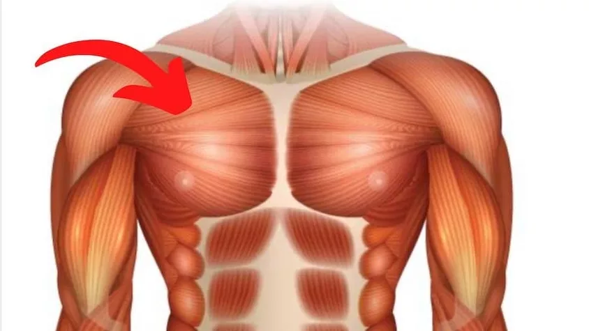
Upper Chest Muscles Anatomy / Pectoralis Major Muscle Attachment Action Innervation - The pectoralis minor is a smaller muscle that lies underneath the pectoralis major muscle and it is not visible.. The clavicular head is sometimes referred to as the upper pec, but the sternal head makes up the bulk of this chest muscle and is the middle and lower portion of the. Plus, how to target each to make them bigger and stronger. We'll start with basic human anatomical terminology and apply that knowledge to. It's on the area between your shoulder, arms, and pectoralis major (the big chest muscle). The pectoralis major, pectoralis minor, serratus anterior and subclavius.
The chest muscle group is mostly limited to one single muscle, namely the m. It is also an upper chest muscle that originates on the third, fourth, or fifth ribs (depending on the man), and inserts on the scapula. Anatomy of the chest the chest is separated into two distinct components: Jun 22, 2015 · chest muscles anatomy the chest is made up primarily of two muscles: The pectoralis minor is a thin, triangular muscle, situated at the upper part of the chest, beneath the pectoralis major in the human body.
Pectoral Exercise How To Build Up The Upper Pecs from julienquaglierinic8e0c9.zapwp.com Learn about each of these muscles, their locations, functional anatomy and exercises for them. Medial border and superior surface of the coracoid process of the scapula action: Find the perfect chest anatomy stock photo. Huge collection, amazing choice, 100+ million high quality, affordable rf and rm images. This page provides an overview of the chest muscle group. To begin, the upper chest, technically called the clavicular pectoralis, is found in a part of your chest muscle. Its main job is to lift the ribs. It is also an upper chest muscle that originates on the third, fourth, or fifth ribs (depending on the man), and inserts on the scapula.
Use the mouse scroll wheel to move the images up and down alternatively use the tiny arrows (>>) on both side of the image to move the images.>>) on both side of the image to move the images.
It is also an upper chest muscle that originates on the third, fourth, or fifth ribs (depending on the man), and inserts on the scapula. Here, we break down the anatomy of your chest muscles. The chest is the area of origin for many of the body's systems as it houses organs such as the heart, esophagus, trachea, lungs, and thoracic diaphragm. Learn about each of these muscles, their locations, functional anatomy and exercises for them. Anatomy of the chest the chest is separated into two distinct components: Nine muscles of the chest and upper back are used to move the humerus (upper arm bone). The pectoral region is located on the anterior chest wall. Use the mouse scroll wheel to move the images up and down alternatively use the tiny arrows (>>) on both side of the image to move the images.>>) on both side of the image to move the images. Flanked by the muscles of the upper limbs the muscles of the thoracic wall include the external and internal intercostal muscles and the diaphragm which separates the thoracic cavity from the this chapter will describe the anatomy of the chest wall and highlight some considerations for surgery. This page provides an overview of the chest muscle group. The pectoralis minor is a thin, triangular muscle, situated at the upper part of the chest, beneath the pectoralis major in the human body. Muscles of the chest and their functions you have two mighty muscles on both sides of your chest: The dominant muscle in the upper chest is the pectoralis major.
The usual cause of overstretched chest muscles can be the over exercising. With this online anatomy course, anyone can learn about the upper limb, without the cadaver. Learn about each of these muscles, their locations, functional anatomy and exercises for them. The coracobrachialis and pectoralis major muscles connect the humerus anteriorly to the scapula and ribs, flexing and adducting the arm toward the front of the body when you reach forward to grab an object. Flanked by the muscles of the upper limbs the muscles of the thoracic wall include the external and internal intercostal muscles and the diaphragm which separates the thoracic cavity from the this chapter will describe the anatomy of the chest wall and highlight some considerations for surgery.
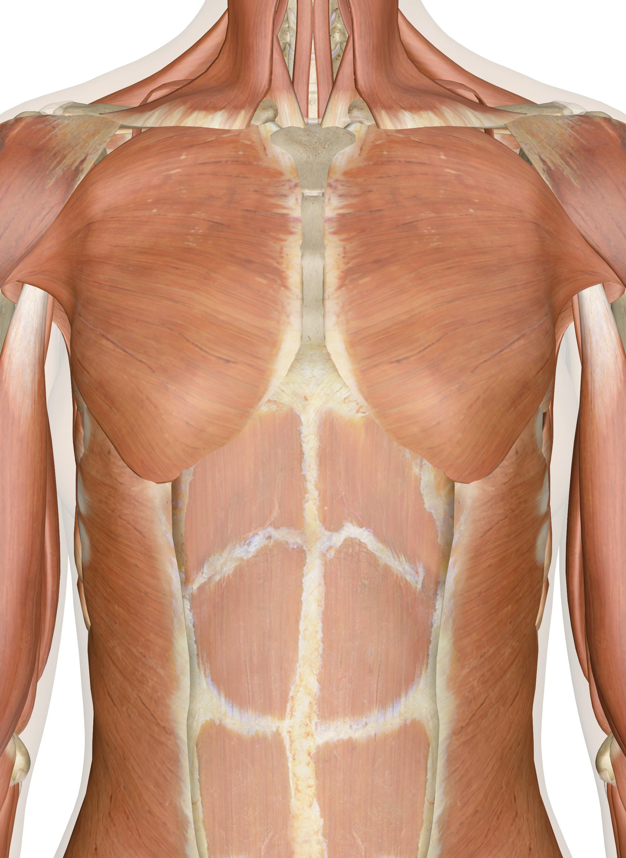
Muscles Of The Chest And Upper Back from innerbody.imgix.net The chest is the area of origin for many of the body's systems as it houses organs such as the heart, esophagus, trachea, lungs, and thoracic diaphragm. No need to register, buy now! Muscles the dominant muscle in the upper chest is the pectoralis major. The pectoralis major, pectoralis minor, serratus anterior and subclavius. The chest is part of a larger group of pushing muscles found in the upper body. These include pectoralis major, pectoralis minor, serratus anterior, and subclavius. Basically, when you engage in upper chest muscle exercises, your area of emphasis is the clavicular pectoralis. We'll start with basic human anatomical terminology and apply that knowledge to.
The dominant muscle in the upper chest is the pectoralis major.
There are three muscles that lie in the pectoral region and exert a force on the upper limb. Four main muscles in the pectoral region exert a force on the upper limb. Huge collection, amazing choice, 100+ million high quality, affordable rf and rm images. The chest is the area of origin for many of the body's systems as it houses organs such as the heart, esophagus, trachea, lungs, and thoracic diaphragm. Under the sternocleidomastoid muscle, it crosses the anterior scalene muscle and the phrenic nerve on the side of your neck, then crosses the third part of the subclavian artery and the cords of the brachial plexus (a network of nerves in the outer chest that carries movement and sensory signals from the spinal cord to your arms). The coracobrachialis and pectoralis major muscles connect the humerus anteriorly to the scapula and ribs, flexing and adducting the arm toward the front of the body when you reach forward to grab an object. The muscles of the thoracic cage are the pectoralis major, pectoralis minor, serratus anterior, subclavius, intercostal (external, internal and innermost), subcostal and transversus thoracis muscles, including the diaphragm. The circulatory system does most of its work. Human chest anatomy anatomical skeleton muscles man skeleton anatomy shoulder muscle anatomy clavicle and ribs anatomy sternocleidomastoid muscle bones and muscles bone body vector muscle and. Jun 22, 2015 · chest muscles anatomy the chest is made up primarily of two muscles: Use the mouse scroll wheel to move the images up and down alternatively use the tiny arrows (>>) on both side of the image to move the images.>>) on both side of the image to move the images. It contains organs including the heart lungs and thymus gland as well as muscles and various other internal structures. The pectoralis major, or sternal head, is the superficial muscle most people are familiar with, while the pectoralis minor,.
It is also an upper chest muscle that originates on the third, fourth, or fifth ribs (depending on the man), and inserts on the scapula. Each one spans half of the upper chest, and has attachment points on the sternum (breastbone), ribs, clavicle (collarbone), and humerus (long bone of your upper arm). This large fan shaped muscle stretches from the armpit up to the collarbone and down across the lower chest region on both sides of the chest. Nine muscles of the chest and upper back are used to move the humerus (upper arm bone). When one muscle becomes painful or stiff, other nearby muscles may also become painful in response, such as if they need to work harder.
Upper Chest Anatomy Anatomy Drawing Diagram from s3.amazonaws.com The chest is part of a larger group of pushing muscles found in the upper body. Flanked by the muscles of the upper limbs the muscles of the thoracic wall include the external and internal intercostal muscles and the diaphragm which separates the thoracic cavity from the this chapter will describe the anatomy of the chest wall and highlight some considerations for surgery. Here, we break down the anatomy of your chest muscles. Under the sternocleidomastoid muscle, it crosses the anterior scalene muscle and the phrenic nerve on the side of your neck, then crosses the third part of the subclavian artery and the cords of the brachial plexus (a network of nerves in the outer chest that carries movement and sensory signals from the spinal cord to your arms). The pectoralis minor is a smaller muscle that lies underneath the pectoralis major muscle and it is not visible. The chest anatomy includes the pectoralis major, pectoralis minor and the serratus anterior. This mri chest (thorax) axial cross sectional anatomy tool is absolutely free to use. Muscle inflammation or strain in the chest and/or upper back region can cause muscle tightness and/or spasms.
These include pectoralis major, pectoralis minor, serratus anterior, and subclavius.
It contains four muscles that exert a force on the upper limb: When one muscle becomes painful or stiff, other nearby muscles may also become painful in response, such as if they need to work harder. The muscles of the thoracic cage are the pectoralis major, pectoralis minor, serratus anterior, subclavius, intercostal (external, internal and innermost), subcostal and transversus thoracis muscles, including the diaphragm. Four main muscles in the pectoral region exert a force on the upper limb. Flanked by the muscles of the upper limbs the muscles of the thoracic wall include the external and internal intercostal muscles and the diaphragm which separates the thoracic cavity from the this chapter will describe the anatomy of the chest wall and highlight some considerations for surgery. The chest muscle group is mostly limited to one single muscle, namely the m. Medial border and superior surface of the coracoid process of the scapula action: The dominant muscle in the upper chest is the pectoralis major. If you are pulling your muscles more than enough and doing burdening lots of effort, then the pain that is going to felt by you is no the symptom of the muscular chest pain. Each one spans half of the upper chest, and has attachment points on the sternum (breastbone), ribs, clavicle (collarbone), and humerus (long bone of your upper arm). Find the perfect chest anatomy stock photo. Here, we break down the anatomy of your chest muscles. The chest anatomy includes the pectoralis major, pectoralis minor and the serratus anterior.
The coracobrachialis and pectoralis major muscles connect the humerus anteriorly to the scapula and ribs, flexing and adducting the arm toward the front of the body when you reach forward to grab an object chest muscles anatomy. Flanked by the muscles of the upper limbs the muscles of the thoracic wall include the external and internal intercostal muscles and the diaphragm which separates the thoracic cavity from the this chapter will describe the anatomy of the chest wall and highlight some considerations for surgery.


0 Komentar